animal cell under microscope 1000x
A drop of blood under the microscope which includes red blood cells white blood cells and platelets. At 40x magnification you will be able to see 5mm.
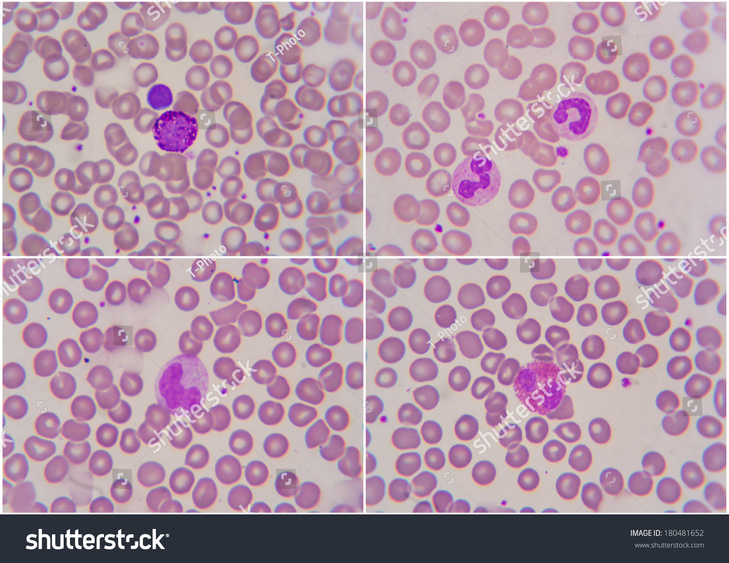
Human Blood Cell Under Microscope 1000x Stock Photo 180481652 Shutterstock
Animal cells usually are transparent and colorless and the thickness of the cell differs throughout the.
. The Cell as a City. Animal Cell Under Microscope. IStock Human Blood Cell Under Microscope 1000x Stock Photo -.
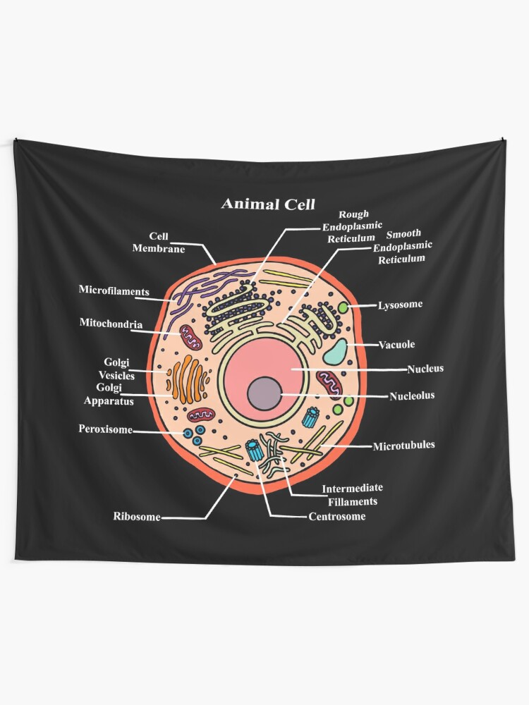
You know Animal cell structure contains only 11 parts out of the 13 parts you saw in the plant cell diagram because Chloroplast and Cell Wall are available only. Using a light microscope one can view cell walls vacuoles cytoplasm chloroplasts nucleus and cell. Animal Cell Under Microscope 1000X Animal Cell Microscope High Res Stock Images Shutterstock - The compound microscope the compound microscope see figure 1.
Can you see cells under a microscope. The air-fixed stained spermatozoa are observed under a bright-light microscope at 400x or 1000x magnification. Transmission electron microscopes are better for the observation.
Mcroscope magnification 400X and 1000X. Great video footage that you wont find anywhere else. Another difference is that plant cells contain one big vacuole whereas animal cells have many smaller ones instead.
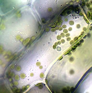
Onion and cheek cells were observed. Moss leaf chloroplasts under microscope 1000x cheek epithelial cells. The compound microscope typically has three or four magnifications - 40x 100x 400x and sometimes 1000x.
40X - 100X - 400X - YouTube from. The magnification of a compound microscope is most commonly 40x 100x 400x and sometimes 1000x. Onion cells under the microscope.
At 1000x magnification you will be able to see 0180mm or 180. What can you see in an animal cell under a light microscope. Cheek cells under the MicroscopeEpithelial cellsstained with methylene bluestained with Gram stainedobservation at magnification of100X400X1000XCheek cells a.
Animal Cell Under Microscope. Onion epidermal cells appear as a. Iii Presence of cell wall.
To identify plant and animal cells you must use a microscope with at least 100x magnification power. Maximum magnification of the brightfield microscope is 100x but modification can readjust the magnification to 1000x which is the optimum magnification of bacterial cells. Animal cell under the microscope.

Aiboully Hd Video Biological Microscope Animal And Plant Blood Cell Analysis Human Health Examination With 7 Inch Screen 1000x Aliexpress Tools

File Frogrbc1000x Jpg Wikipedia
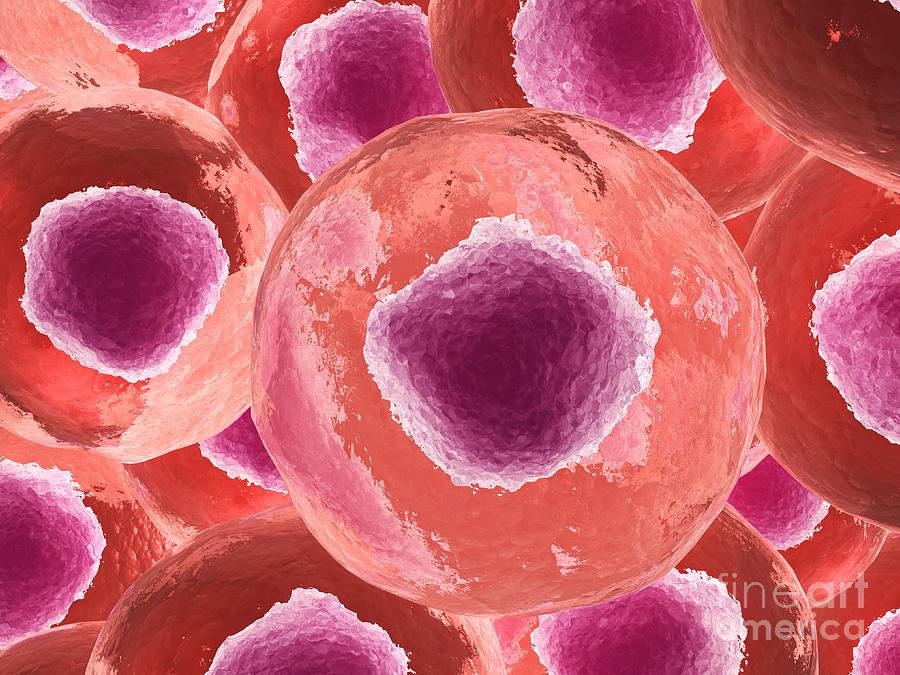
Microscopic View Of Animal Cell Digital Art By Stocktrek Images Fine Art America

Structure Of Animal Cell And Plant Cell Under Microscope Diagrams
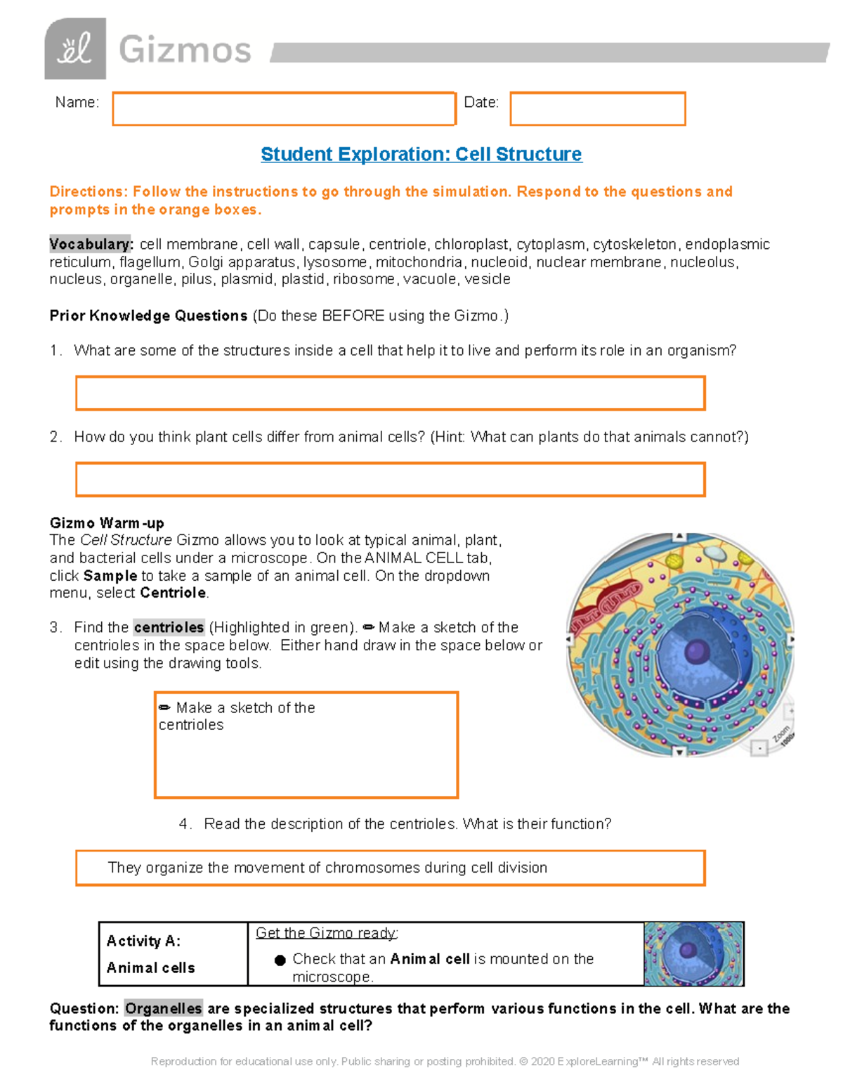
Copy Of Cell Structure Se Aaaaaaaaaaaaaaaaaaaaaaaaaaa Aaaaaaaaaaaaaaaaaaaaaaaaaaa Studocu
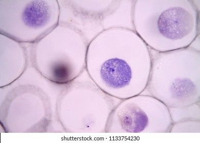
Mitosis Animal Cell Under Microscope Stock Photo 1133754230 Shutterstock
Solved Bio 101 Lab 03 Microscopy And Cells Notification Objectives Course Hero
What Are The Visible Plant Animal Cell Organs On Light Microscope Quora

Cyanobacteria Photos
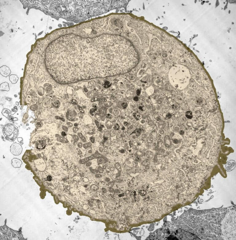
What Does An Animal Cell Look Like Under An Electron Microscope Quora
Cell Structure
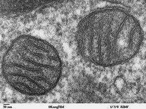
Mitochondrion Wikipedia

Haines Educational Science Catalogue 2018 By Haines Educational Issuu

Anatomy And Physiology Of Animals The Cell Wikieducator
Amazing 27 Things Under The Microscope With Diagrams
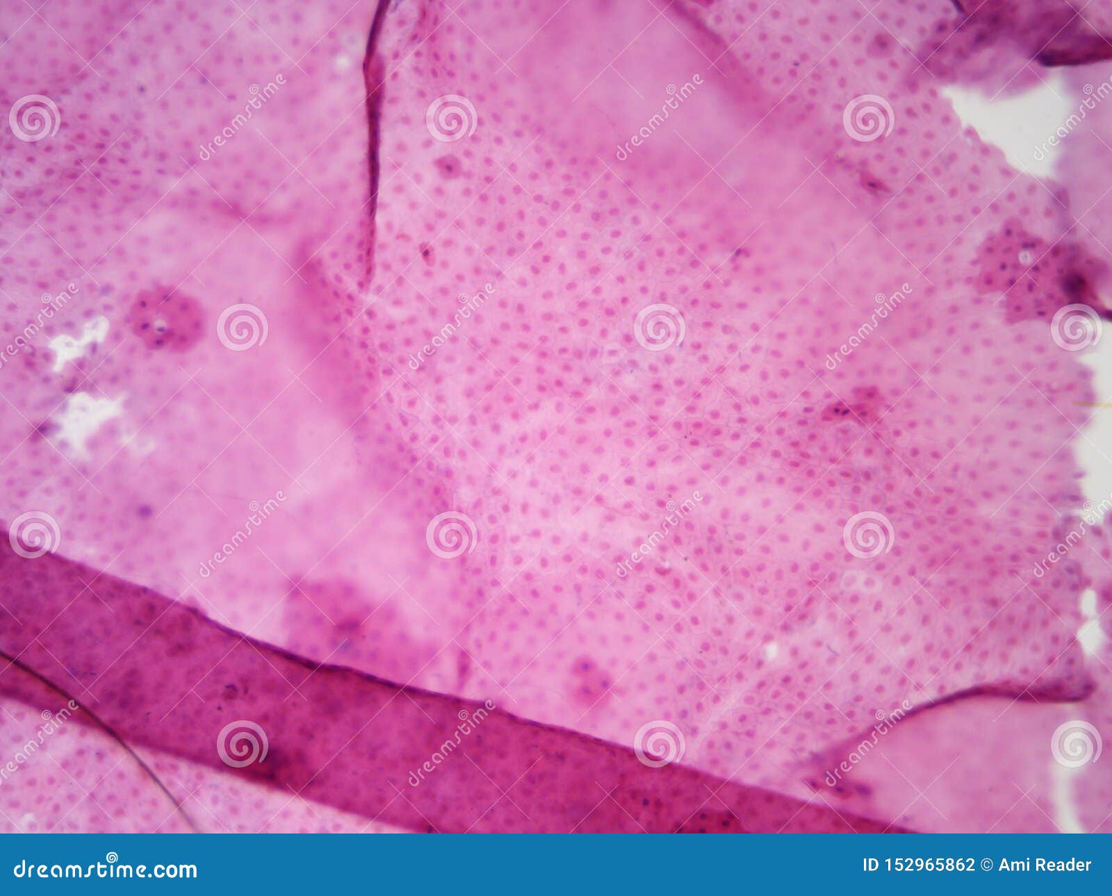
Typical Animal Cell Center 100x Stock Photo Image Of 100x School 152965862
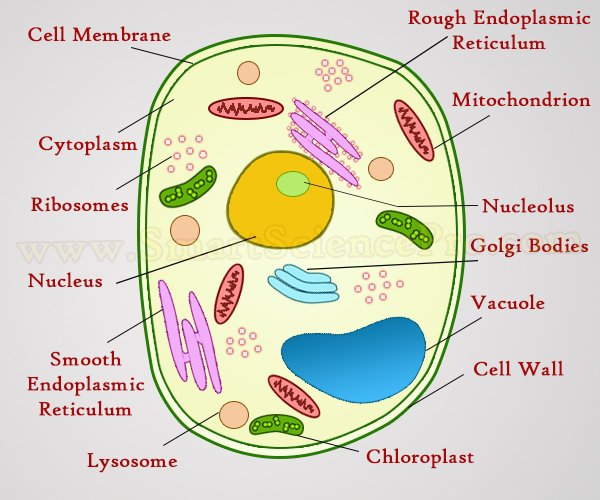
Structure Of Animal Cell And Plant Cell Under Microscope Diagrams

Prophase

Human Blood Cell Under Microscope 1000x Stock Photo 180481652 Shutterstock